
Why Did My NAD+ Turn Yellow? Colour, Stability and Storage
A pale yellow NAD+ solution sits at or beyond the edge of what reagent datasheets describe: most call freshly dissolved NAD+ clear and colourless, and one specification allows colourless to faintly yellow. What the colour does not reveal is what caused the tint or how much NAD+ is left. NAD+ is not a peptide but a dinucleotide coenzyme, and it is approved as a medicine only in China, as an injection. This page explains the documented chemistry and marks clearly where that chemistry stops.
Is NAD supposed to be yellow? What colour NAD+ is at the start
Is NAD plus yellow when it is freshly dissolved? Mostly not, in the descriptions that manufacturers of laboratory reagents give: whether it is called NAD, NAD+ or NAD plus, they describe a fresh solution as colourless, and one specification allows at most a faint yellow. NAD+ (nicotinamide adenine dinucleotide, 663.43 g/mol) is two nucleotides joined by a phosphate bridge. It contains no amino acids, which is why the question "is NAD peptide yellow" rests on a mislabel: the molecule is sold beside peptides, but its colour follows nucleotide chemistry.
NAD+ powder color
The dry substance is described as a white to off-white lyophilised (freeze-dried) solid in one datasheet, as off-white in another, and as a crystalline solid in a third. One specification sheet allows a range of "white to yellow" for the powder, so a faintly cream or yellowish powder can still fall within a manufacturer's specification. The powder is hygroscopic, meaning it draws water from the air, and datasheets store it dry.
NAD+ liquid color
Dissolved in water, NAD+ is described as giving "a clear, colorless solution". A second specification sheet records the same for a 1 percent solution. A third, for the sodium salt of NAD+ at 50 mg/ml, allows a solution that is "clear, colorless to faintly yellow". NAD+ absorbs ultraviolet light at 260 nm, which the eye cannot see, and it does not absorb at 340 nm.
The reduced form, NADH, behaves differently. NADH is formed when NAD+ takes up a hydride ion, a step that enzymes called dehydrogenases carry out. Its altered ring (a dihydropyridine ring) absorbs at 340 nm as well as at 260 nm, and one NADH datasheet records weaker absorption at 365 nm too. One manufacturer accordingly describes NADH powder as "white to slightly yellowish" and its solution as "clear, colourless to slightly yellowish". NAD+ and NADH are not interchangeable names: they are two different substances with different colour descriptions.
Why NAD turned yellow: the chemistry in water
NAD+ is stable as a dry solid and much less stable in water. The literature describes several routes by which it changes, and each depends on pH, temperature, light and time.
Hydrolysis. In water, the bond that links the nicotinamide ring of NAD+ to its sugar breaks, releasing nicotinamide and ADP-ribose. A kinetic study describes NAD+ as undergoing "pH-independent and base-catalyzed hydrolysis", with the pH-independent route dominating at pH 6 and below. Work on heat breakdown confirmed nicotinamide and ADP-ribose as the predominant products.
Alkaline conditions. NAD+ is destroyed faster as a solution becomes more alkaline. One datasheet calls NAD+ solutions "very labile in alkaline solutions, especially in the presence of phosphate, maleate, or carbonate", and another states that the product "will decompose at alkaline pH". NADH shows the opposite pattern and breaks down faster in acid. In strong alkali, NAD+ turns into a highly fluorescent product, which laboratories use to measure it. The colour of that product is not stated in the sources reviewed for this page.
Heat and light. Solutions "are rapidly degraded upon heating" and "are also sensitive to light", according to the same datasheet.
Reducing sugar chemistry. ADP-ribose, one hydrolysis product, contains a reducing sugar, a type of sugar that can react with amino groups. With proteins it forms early glycation products and later fluorescent compounds; this kind of glycation is the opening stage of the Maillard reaction, which goes on to form so-called advanced glycation end products. Those studies used proteins or amines as reaction partners. Whether this chemistry produces a yellow tint in a solution of NAD+ alone could not be identified in the sources reviewed for this page.
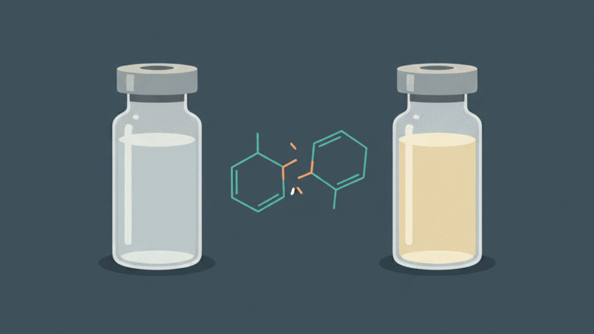
The honest summary for a solution where NAD+ has a yellow tint is therefore limited. The documented changes are hydrolysis, alkaline breakdown and heat and light sensitivity. NADH, which datasheets describe as slightly yellowish, forms from NAD+ when an enzyme transfers a hydride; whether NADH forms in a stored NAD+ solution could not be established from the sources reviewed for this page. Which of these routes, if any, is responsible for a particular yellow tint could not be identified in the sources reviewed for this page. That matches the wider rule for discolouration described on the peptide storage page: yellowing is often associated with oxidation or Maillard chemistry, but it does not reveal which reaction took place or how far it went.
NAD+ vial color, syringe color and the thickness of the liquid
The same liquid can look different depending on the container. The Beer-Lambert law, as defined by IUPAC, states that the absorbance of light is proportional to the concentration of the absorbing substance and to the distance the light travels through it. A more concentrated solution therefore looks darker, and so does a wider column of the same solution. A narrow syringe barrel gives a short path, a vial viewed through its full width a longer one, and looking down through a filled vial from above longer still.
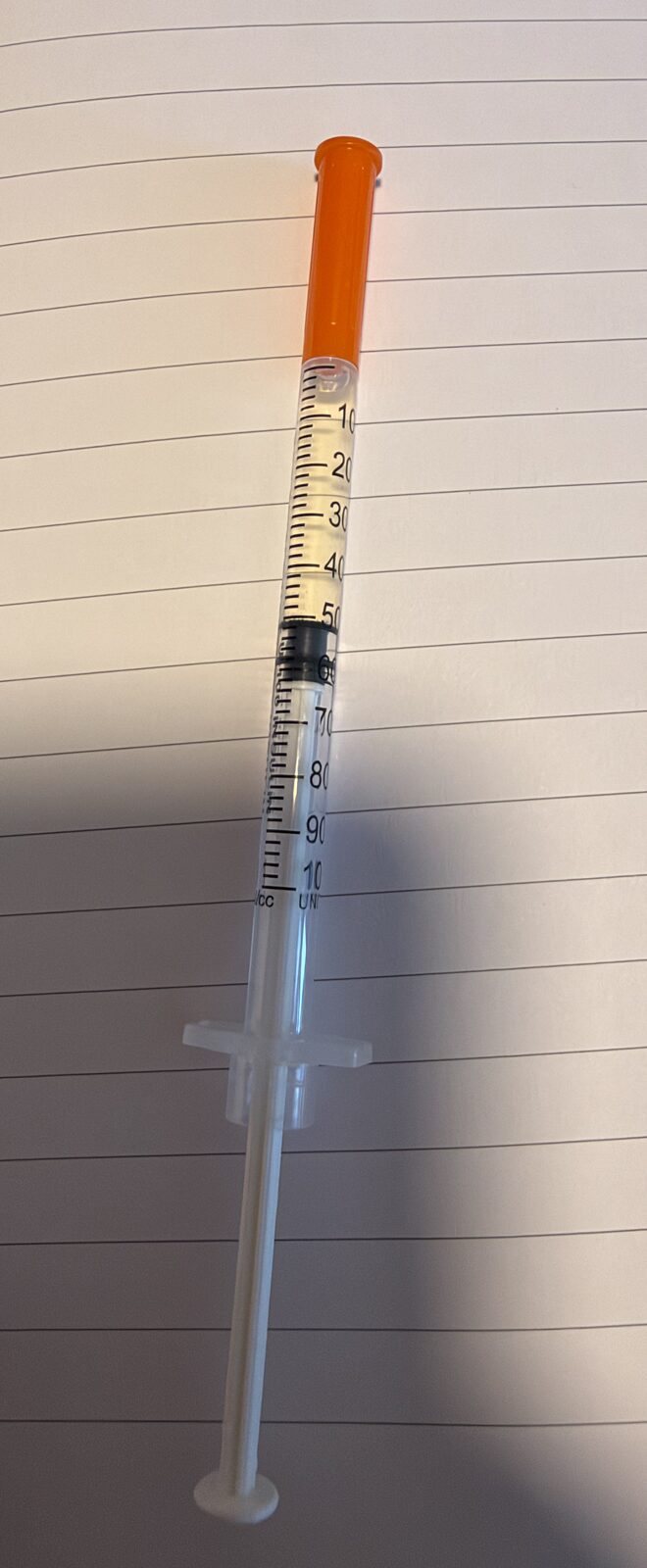
Editorial photo of a pale yellow NAD+ solution in an insulin syringe. The colour on its own says nothing about how much NAD+ the liquid contains or how pure it is.
Concentration itself is plain arithmetic: the mass of substance divided by the volume of liquid. The NAD+ reconstitution calculator works through that ratio, and the reconstitution basics explain the units. A small volume of water over the same mass gives a more concentrated solution, which on the Beer-Lambert relationship absorbs more light in the same container.
How fast NAD+ degrades in solution
Published figures come from laboratory buffers and controlled temperatures, not from vials kept at home, and they are not directly transferable. With that limit, they show how strongly pH, temperature and buffer decide the pace.
| Condition | Molecule | Reported result | Source |
|---|---|---|---|
| Tris buffer, pH 8.5, 19 °C, 43 days | NAD+ (2 mM) | 260 nm signal fell by 4 percent | Wolfe et al. 2024 |
| HEPES buffer, pH 8.5, 19 °C, 43 days | NAD+ (2 mM) | Degraded nearly entirely | Wolfe et al. 2024 |
| Sodium phosphate buffer, pH 8.5, 19 °C, 43 days | NAD+ (2 mM) | Degraded at a rate similar to NADH in the same buffer | Wolfe et al. 2024 |
| Tris buffer, pH 8.5, 19 °C vs 25 °C | NADH (2 mM) | 4 vs 11 µM lost per day; over 90 vs 75 percent left after 43 days | Wolfe et al. 2024 |
| HEPES buffer, pH 8.5, 19 °C vs 25 °C | NADH (2 mM) | 18 vs 51 µM lost per day | Wolfe et al. 2024 |
| Sodium phosphate buffer, pH 8.5, 19 °C vs 25 °C | NADH (2 mM) | 23 vs 34 µM lost per day | Wolfe et al. 2024 |
| Aqueous solution, 10 mg/ml, 2 to 8 °C, 4 weeks | NAD+ | At least 99 percent remaining | Roche datasheet |
| Stock solution, pH 2 to 7, 4 °C | NAD+ | Stable for up to 1 month | Calbiochem datasheet |
| Neutral or slightly acidic solution, 0 °C | NAD+ | Stable for at least 2 weeks | Sigma-Aldrich datasheet |
| Solution pH 2 to 6, single-use portions, -70 °C | NAD+ | Stable for at least 6 months | Sigma-Aldrich datasheet |
| 50 mM Tris, pH 6.5 (measured at 85 °C), 85 °C | NAD+ (2 mM) | Half-life 24.2 minutes; below 25 percent left after 60 minutes | Hachisuka et al. 2017 |
| 0.1 M Tris, pH 7.60 (measured at 25 °C), 100 °C | NAD+ | Half-life about 10 minutes | Hachisuka et al. 2017, citing earlier work |
µM stands for micromoles per litre; the half-life is the time after which half of the starting amount is gone. The authors of the 43-day study describe their NAD+ results as qualitative, because breakdown products of NADH can also absorb at 260 nm, the wavelength used to follow NAD+. They also note a "clear red shift" of the 260 nm band as NAD+ degraded.
NAD slight yellow tint: what it does and does not tell you
Finding NAD+ slightly yellow in a vial says that the solution now absorbs some visible light that a fresh NAD+ solution did not. It does not say how much NAD+ remains. In the 43-day study, an NAD+ solution in HEPES buffer degraded nearly entirely, and that loss was measured through ultraviolet absorbance at 260 nm, which the eye cannot see; the study did not describe the visible colour of its solutions. Colour and remaining content are therefore separate observations, and a faint tint does not by itself establish either a small or a large loss.
Colour is not a measure of strength or purity. Content and purity are what laboratory methods measure, for example HPLC (high-performance liquid chromatography, which separates and quantifies each component) or the enzymatic assays named in reagent datasheets. A batch certificate of analysis records those results for the material as supplied, not for a solution after it has been made up and stored. When NAD looks yellow in one container and paler in another, concentration and path length explain much of the difference, but neither tells you what the tint is made of.
Storage conditions described in datasheets and studies
The published conditions are consistent in direction, even though they come from laboratory reagents rather than from any product for human use.
- Dry powder: stored at -20 °C in several datasheets and at 2 to 8 °C in another, kept dry because the solid is hygroscopic. One manufacturer gives a stability of at least 4 years at -20 °C for the solid.
- pH: neutral to slightly acidic solutions are described as the more stable; alkaline solutions are described as labile.
- Temperature: in the 43-day study, a rise from 19 °C to 25 °C raised the NADH loss rate from 4 to 11 µM per day in Tris buffer and from 18 to 51 µM per day in HEPES buffer, and heating is described as causing rapid degradation.
- Light: NAD+ solutions are described as light-sensitive, and NADH datasheets specify protection from light.
- Time: one manufacturer advises against keeping an aqueous NAD+ solution for more than one day, while others report at least 2 weeks at 0 °C or at least 4 weeks at 2 to 8 °C for specific solutions.
- Freezing: one ready-made 50 mM NAD+ solution is stored at -20 °C, with repeated freezing and thawing avoided and -80 °C for storage beyond 30 days.
These are the conditions recorded for laboratory reagents. They are described here as published findings, not as a handling procedure. Further background sits in peptide storage, peptides at room temperature and bacteriostatic water.
What pharmacopoeias say about visible change
NAD+ injection color and the pharmacopoeial standard
Pharmacopoeias set binding quality standards for medicines. They are relevant here because they describe what an injectable solution looks like when it meets the standard. The European Pharmacopoeia states that "solutions for injection, examined under suitable conditions of visibility, are clear and practically free from particles." The United States Pharmacopeia requires an inspection process designed so that every lot of parenteral preparations, meaning medicines given by injection or infusion, "is essentially free from visible particulates", and the Japanese Pharmacopoeia uses comparable wording.
Colour is handled through a separate test method. Under European Pharmacopoeia method 2.2.2, reproduced in the International Pharmacopoeia, a liquid is compared with reference solutions in the brown, brownish-yellow, yellow, greenish-yellow and red range. It counts as colourless if it looks like water or is not more intensely coloured than the most dilute brown reference solution, B9. The method fixes the geometry: 2.0 mL in a 12 mm tube viewed from the side against a white background, or a 40 mm deep layer viewed from above. On the Beer-Lambert relationship described earlier, the depth of liquid affects how intense a colour appears. The permitted colour is set by the monograph for each individual product.
No pharmacopoeial monograph for an NAD+ injection could be identified in the sources reviewed for this page. That fits the approval picture: an injectable NAD+ product is approved only in China, and none is approved in Europe or the United States. The European and American standards above therefore describe approved medicines, not NAD+ sold outside that system.
Common questions
Is NAD supposed to be yellow?
Not as a rule. Reagent datasheets describe NAD+ powder as white to off-white or off-white, with one specification allowing white to yellow. They describe a fresh solution in water as clear and colourless, and one specification allows colourless to faintly yellow. A yellow tint beyond that faint range departs from these descriptions, but the colour alone does not identify its cause.
Is NAD clear or yellow?
Freshly dissolved NAD+ is described in datasheets as a clear, colourless solution, and one specification allows colourless to faintly yellow. The reduced form, NADH, is a different substance: one datasheet describes its solution as colourless to slightly yellowish. NADH also absorbs light at 340 nm, where NAD+ does not.
Is NAD peptide yellow?
NAD+ is not a peptide. It is a dinucleotide, a coenzyme built from two nucleotides joined by a phosphate bridge, with a mass of 663.43 g/mol and no amino acids. It is sold beside peptides, which is where the label comes from. Its colour behaviour follows nucleotide chemistry, not peptide chemistry.
My NAD is yellow: what does a slight yellow tint mean?
It means the solution now absorbs some visible light that a fresh NAD+ solution does not. The documented ways NAD+ changes in water are hydrolysis into nicotinamide and ADP-ribose, faster with heat and in alkaline solution, and sensitivity to light. Which of these, if any, produces a yellow tint could not be identified in the sources reviewed for this page.
Why does NAD look yellow in one container and paler in another?
How much light a solution absorbs rises with the concentration and with the distance the light travels through it, a relationship known as the Beer-Lambert law. A narrow syringe barrel gives a short path, a wide vial a longer one, so the same liquid can look darker in the vial. Pharmacopoeial colour tests specify the tube size and the viewing direction.
Does NAD+ need to be refrigerated?
Reagent datasheets store the dry powder at -20 °C or at 2 to 8 °C, kept dry because it is hygroscopic. For solutions they report at least 99 percent remaining after 4 weeks at 2 to 8 °C for one aqueous solution, stability for at least 2 weeks at 0 °C for neutral or slightly acidic solutions, and rapid degradation on heating.
Does a yellow tint show how much NAD+ is left?
No. Colour is not a measure of content. In a 43-day study, an NAD+ solution in one buffer lost nearly all of its NAD+; the loss was measured by ultraviolet absorbance, not by eye, and the study did not describe the visible colour. Content and purity are established by laboratory analysis, such as HPLC or an enzymatic assay, not by looking at the vial.
Is NAD+ approved as a medicine?
Only in China. There an NAD+ injection is approved for the supportive treatment of low white-cell counts, coronary heart disease and myocarditis. No approval was found in the United States, the European Union, the United Kingdom, Canada, Australia or Switzerland. Reagent manufacturers sell NAD+ for laboratory use, and one datasheet states that it is not for drug use.
Sources
- Sigma-Aldrich (2006). β-Nicotinamide adenine dinucleotide, Product Number N8285, Product Information. Sigma-Aldrich. https://www.sigmaaldrich.com/deepweb/assets/sigmaaldrich/product/documents/668/557/n8285pis.pdf
- Merck / Calbiochem (2026). NAD+, Free Acid, product 481911: properties and preparation note. Merck KGaA. https://www.sigmaaldrich.com/US/en/product/mm/481911
- Roche (2026). NAD Grade I, free acid: specification and storage of working solution. Roche Diagnostics via Sigma-Aldrich. https://www.sigmaaldrich.com/US/en/product/roche/nad100ro
- Roche CustomBiotech (2026). NADH, Grade II, disodium salt: appearance, solution and stability. Roche Diagnostics. https://custombiotech.roche.com/global/en/products/cb/nadh-grade-ii-322141.html
- Roche (2026). NADH Grade II, disodium salt: absorbance coefficients and storage. Roche Diagnostics via Sigma-Aldrich. https://www.sigmaaldrich.com/US/en/product/roche/10128023001
- Thermo Fisher Scientific (2026). beta-Nicotinamide adenine dinucleotide hydrate, 98+%, catalogue 124532500, Product Specification. Thermo Fisher Scientific. https://assets.thermofisher.com/chem-specs-pdf/retrievePdf?countryCode=cn&erp_type=CN_E1_LCD&rootSku=12453&sku=124532500
- Cayman Chemical (2022). NAD+ (free acid), Item No. 16077, Product Information. Cayman Chemical. https://cdn.caymanchem.com/cdn/insert/16077.pdf
- New England Biolabs (2026). β-Nicotinamide adenine dinucleotide (NAD+), B9007, product notes. New England Biolabs. https://www.neb.com/en-us/products/b9007-beta-nicotinamide-adenine-dinucleotide-nad
- Wolfe KD, Alahuhta M, Himmel ME, Bomble YJ, Jennings GK, Cliffel DE (2024). Long-Term Stability of Nicotinamide Cofactors in Common Aqueous Buffers: Implications for Cell-Free Biocatalysis. Molecules 29(22):5453. https://pmc.ncbi.nlm.nih.gov/articles/PMC11597533/
- Hachisuka S-I, Sato T, Atomi H (2017). Metabolism Dealing with Thermal Degradation of NAD+ in the Hyperthermophilic Archaeon Thermococcus kodakarensis. Journal of Bacteriology 199(19):e00162-17. https://pmc.ncbi.nlm.nih.gov/articles/PMC5585711/
- Bull HG, Ferraz JP, Cordes EH, Ribbi A, Apitz-Castro R (1978). Concerning the mechanism of the enzymatic and nonenzymatic hydrolysis of nicotinamide nucleotide coenzymes. Journal of Biological Chemistry 253(14):5186-5192. https://pubmed.ncbi.nlm.nih.gov/209029/
- Iverson D, DeChatelet LR, Spitznagel JK, Wang P (1977). Comparison of NADH and NADPH oxidase activities in granules isolated from human polymorphonuclear leukocytes with a fluorometric assay. Journal of Clinical Investigation 59(2):282-290. https://pmc.ncbi.nlm.nih.gov/articles/PMC333358/
- Jacobson EL, Cervantes-Laurean D, Jacobson MK (1997). ADP-ribose in glycation and glycoxidation reactions. Advances in Experimental Medicine and Biology 419:371-379. https://pubmed.ncbi.nlm.nih.gov/9193679/
- Cervantes-Laurean D, Minter DE, Jacobson EL, Jacobson MK (1993). Protein glycation by ADP-ribose: studies of model conjugates. Biochemistry 32(6):1528-1534. https://pubmed.ncbi.nlm.nih.gov/8431431/
- IUPAC (2025). Beer–Lambert law. Compendium of Chemical Terminology (Gold Book), version 5.0.0. https://doi.org/10.1351/goldbook.B00626
- World Health Organization (2017). The International Pharmacopoeia, 1.11 Colour of liquid, including 1.11.2 Degree of coloration of liquids reproduced from the European Pharmacopoeia (method 2.2.2). WHO. https://digicollections.net/phint/pdf/b/7.1.11.1.11-Colour-of-liquid.pdf
- Liu F, Hutchinson R (2024). Visible particles in parenteral drug products: A review of current safety assessment practice. Current Research in Toxicology 7:100175. https://doi.org/10.1016/j.crtox.2024.100175
- PubChem (2026). Compound summaries CID 5892 (nadide, NAD+) and CID 439153 (NADH). National Center for Biotechnology Information. https://pubchem.ncbi.nlm.nih.gov/compound/5892
- Zhang Q, Ames JM, Smith RD, Baynes JW, Metz TO (2009). A perspective on the Maillard reaction and the analysis of protein glycation by mass spectrometry: probing the pathogenesis of chronic disease. Journal of Proteome Research 8(2):754-769. https://pubmed.ncbi.nlm.nih.gov/19093874/
- Sigma-Aldrich (2026). β-Nicotinamide adenine dinucleotide sodium salt, Product Number N0632, product specification: solubility in water 50 mg/ml, clear, colorless to faintly yellow. Sigma-Aldrich. https://www.sigmaaldrich.com/US/en/product/sigma/n0632
- DXY drug database (2026). Label of Coenzyme I for Injection (注射用辅酶Ⅰ), Kaifeng Knature Pharmaceutical, approval numbers H41024721 and H41025094, label approved 8 December 2010. https://drugs.dxy.cn/drug/C0e3Iui9RyaWHKhCVVQQCw==
This page describes colour, chemistry and published storage data only. For a specific batch, the authoritative record of identity, content and purity is the manufacturer's certificate of analysis or an independent laboratory analysis; questions about health or any use of a substance belong with a pharmacist or physician.